

High temperature and density plasma emit in electromagnetic spectra, which includes X-rays. These X-rays hold very important information regarding the plasma interior as well as the plasma boundary. Temporal monitoring of X-ray emission serves a wide range of physical phenomena which assists in better understanding of the tokamak plasma. MHD activities, disruptions, plasma start-up, identification of the magnetic axis, plasma boundary, electron temperature are some of the important physics objectives for which the X-ray diagnostic are designed. X-ray emission ranges between a few hundreds of eV to a few tens of MeV. Tokamak X-rays are majorly divided into two groups Soft X-ray (~800eV -30keV) and Hard X-rays (~100 keV to a few MeV).
Imaging & X-ray
Overview
Imaging & X-ray
Experiments
1.Soft X-ray (SX) Diagnostic (~800 eV - ~30 keV)
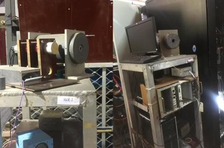
A Soft X-Ray tomographic system is intended to perform the tomographic reconstruction for the identification of the equilibrium emission profile as well as the realization of the magnetic islands available within the plasma, employing AXUV linear photodiode detector array. The diagnostic is a perpendicular viewing (perpendicular to BT) system consisting of 4 arrays, having 16 individual channels. Presently one array is operational, the other one is designed, fabricated and ready for operation and the remaining ones are under development. Along with the tomographic reconstruction the SXR diagnostic is also employed for electron temperature estimation, via the foil method. To facilitate the same an additional array is placed with an extra filter for the determination of the electron temperature. A Soft X-Ray monitor is a single channel-wide Field of View (FOV) x-ray detector system that gives an overall Soft X-ray emission pattern in temporal space. A Silicon Barrier Detector (SBD) is used for this purpose because it has a large detection (collection) area, which helps satisfy the requirement for a wide FOV.
2.Hard X-ray (HX) Diagnostic (~100 keV - ~10 MeV)
The hard X-rays, HX, are produced by the runaway electrons (RE). RE is the most undesirable electron population for any given tokamak plasma, and identification/ subsequent mitigation is a principal requirement for any successful tokamak plasma operations. The two HX monitors are the tangential viewing wide FOV NaI-based scintillator which monitors the HX emission in the temporal space. The HX burst is one of the key pieces of information which is obtained from these systems.
The Hard X-ray Spectrometer system is LaBr (1.5 inches x1.5 inch) scintillator detector based spectrometer, which is again a wide FOV single-channel system. This offers the temporal evolution of the HX spectra for any given tokamak plasma discharge. The diagnostic main objective is to give the runaway electron temperature.
3.High Resolution Visible Imaging Diagnostic (HR-VID)
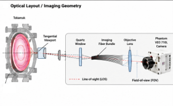
The High Resolution Visible Imaging Diagnostic (HR-VID) is an advanced optical diagnostic system installed on the ADITYA-U and SST-1 tokamak for high-speed visualization of plasma behaviour. The diagnostic provides time-resolved, two-dimensional (2D) imaging of the plasma, enabling detailed studies of plasma evolution, edge phenomena, and transient events during tokamak discharges.
The system is installed at a tangential mid-plane viewing configuration and comprises a precision C-mount objective lens, a wound imaging fiber bundle, a relay lens, and a high-speed Phantom VEO 710 camera. The diagnostic captures images of the poloidal cross-section of the plasma with a temporal resolution in the range of (1µs to 30ms). The Phantom VEO 710 camera is capable of recording images at frame rates of up to 7,000 frames per second at full sensor resolution, making it suitable for investigating evolving plasma phenomena.
Image acquisition is synchronized with the tokamak control system through a hardware trigger generated at the start of each plasma discharge. The camera records the complete plasma shot, temporarily storing the acquired image sequence in its on-board high-speed memory. Following the discharge, the data are transferred to the dedicated Data Acquisition (DAQ) computer via Ethernet using the Phantom Camera Control (PCC) software, which also provides comprehensive control over camera operation and acquisition parameters.
Division Head

Section Head

Team Members